An Enhanced System for Pencil Beam Characterization
Background
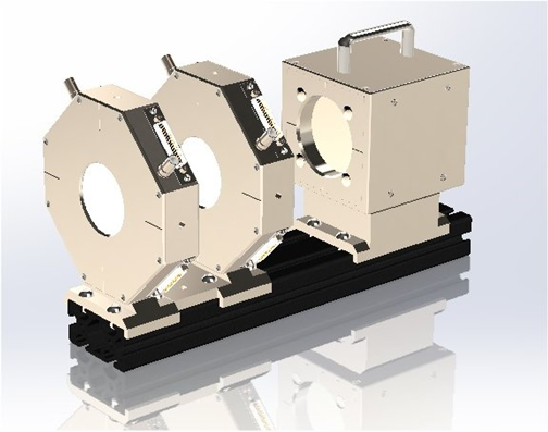
The rapid development and introduction of pencil beam scanning for particle therapy raises new challenges for system commissioning and routine quality assurance. A beam diagnostic system was described at PTCOG 53 which allowed the shape, trajectory, divergence and current of a pencil beam to be simultaneously determined [1]. The system is now being enhanced by the addition of a multilayer Faraday collector (MLFC) [2] which provides beam energy and energy spread information as well as total beam current.
Energy validation is required for light ion therapy for compliance with IEC 60601-2-64 [3]. Potential problems that are detectable with a MLFC are setting the wrong energy, energy degradation due to beam strike along the beamline and excess energy spread due to incorrect energy analyzer setup.
The use of the multilayer Faraday collector was explored by Gottschalk at the Harvard Cyclotron Laboratory [2], and has been used by other groups and manufacturers [3,4,5]. The MLFC can be used as a primary standard, because the accuracy of determination depends in principle only on well-established stopping powers and knowledge of material composition and density. MLFC data has in fact been used to validate Monte-Carlo simulations [6,7]. In contrast to the more complex multi-layer ionization chamber, it measures beam charge directly rather than ionization dose.
We have built a MLFC, suitable for routine manufacture, and done initial testing with proton beams at Massachusetts General Hospital. This has proven that the construction method is sound and that absolute beam energy measurement is feasible. There is also indication of useful sensitivity to beam energy spread.
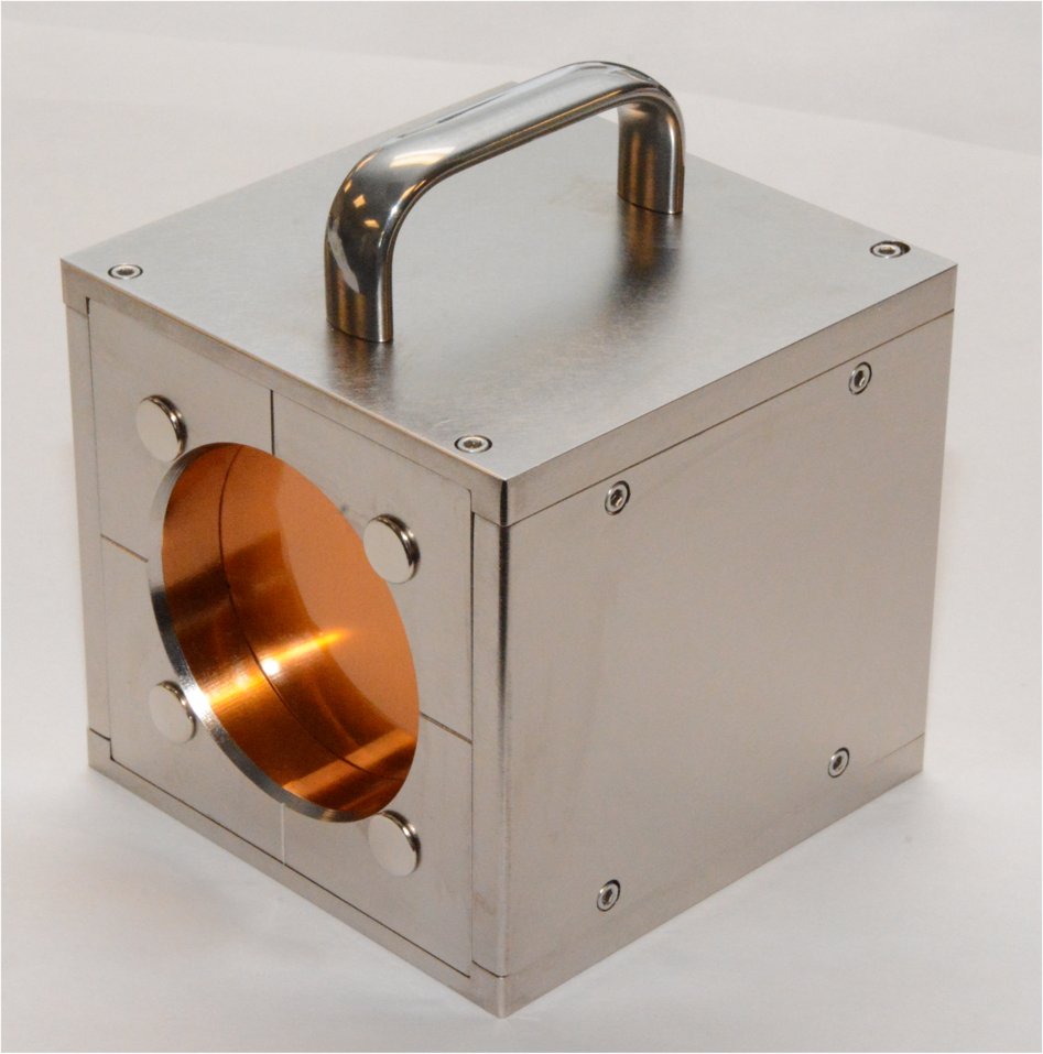
Prototype MLFC assembly with 128 layers of 0.0508 mm copper, suitable for proton beam energies up to 250 MeV. The assembly weighs 5.6 kg (12.2 lb) and measures 120 x 120 x 145 mm. The sheets are fully screened inside the housing and the signals are brought out on two VHDCI68 connectors. The useful area is 72 mm diameter, sufficient for the beam size of most PBS systems.
Method
Much of the prior work using MLFCs has relied on careful manual assembly of bespoke devices. Our objective here is to render the device suitable for routine production while providing the best possible absolute accuracy.
The first version of the device has been built to cover the full proton energy range of interest for particle therapy in the body, 50 to 250 MeV, for general quality assurance. Pure copper is used as the stopping material, to remove uncertainty about composition. The copper sheets were water-jet cut at 25 µm precision to a very accurately known area. Each sheet included a tab to allow a direct solder connection to a PCB which routed the connections to one of two 68-way connectors. This removed the need for individual wired connections. The sheets were manufactured with a better than 1% RMS tolerance on thickness variation.. Each sheet was weighed on a precision balance, giving the areal density in g cm-2 to four significant figures. The sheets are electrically isolated with thin sheets of polyimide film. The effective areal density of each layer is slightly increased accordingly. Gottschalk [2] has explained how the beam charge is always measured, even if the ion or a secondary electron happens to stop in a dielectric layer. The SRIM Monte-Carlo code was used to calculate the longitudinal distribution in g cm-2 of proton stopping in pure copper at multiple energies. Using the measured areal densities of the individual layers, we create a table of range in the MLFC measured in sheet number (representing the longitudinal center of the sheet) as a function of proton energy in 1 MeV steps. Given an unknown beam we fit a Gaussian curve to the end of range peak to get a range expressed in fractional sheet number. Then we simply interpolate the MLFC-specific range table to obtain the beam energy.
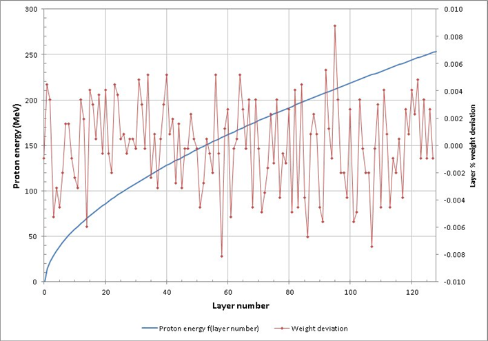
Relationship between MLFC layer number and proton energy for the prototype MLFC which is stored as a device-specific table. The weight deviation curve illustrates the corrections applied in the creation of the table.
Because there is no physical gain in an MLFC, a sensitive electrometer with low background offset is required to collect the signals. The I128 electrometer [8]. was used for the tests described here. The QA procedure can generally allow several seconds for the signal to be integrated to give sufficient signal to noise ratio.
Monte-Carlo calculations predict the amount of straggle in the range of a mono-energetic zero divergence beam. Real beams have some energy spread, the amount depending on the type of accelerator, and some divergence, generally small for PBS proton therapy systems. The measured spread in the range is the convolution of range straggling and energy spread; the beam energy spread can in principle be de-convolved.
Results
The MLFC was exposed to proton Beams at the Francis H Burr Proton Therapy Center at Massachusetts General Hospital. The beam energies on the system are well-characterized. A full energy beam was measured, a full energy beam passed through a Lucite degrader and a lower energy beam created by the degrader/analyzer system on the accelerator. The signals were integrated for about ten seconds with 100 msec resolution.
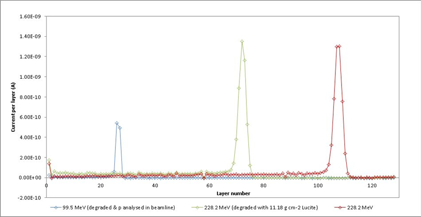
Raw data for three different beam energy conditions. The low-energy tails are the result of nuclear scattering secondaries.
At the lowest energy the energy selector slits were closed in steps 40 - 20 - 10 -5 mm to see if the effect on energy spread was measurable. Only the initial step was directly visible, but curve fitting did reveal a linear trend. If we make the conservative assumption the smallest slit gave an energy spread that the MLFC could no longer resolve from the range straggling, then this implies an energy spread of 0.83 MeV or 0.83% with the 40 mm setting. An energy spread resolution of better than 0.2% is indicated by the result.
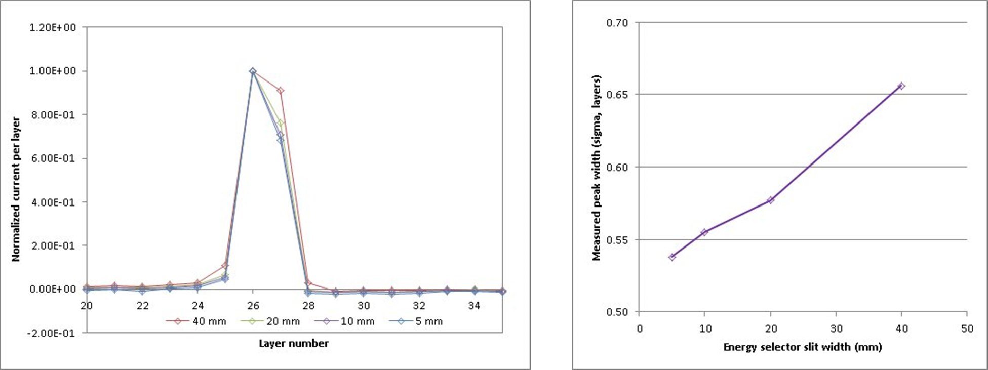
Detection of effect of energy selector slit width
Further work
The MLFC will be exposed to a more complete range of energies from different accelerator types, and the results will be compared to measured range in water. A variant using thinner materials will be optimized for low energy measurements. The MLFC will be integrated with the pixilated ionization chamber telescope to give a single device which measures all critical beam parameters for pencil beam scanning.
Acknowledgements
We are grateful as ever to Ethan Cascio for his efficient assistance taking data at MGH, and to Dr Jay Flanz for permission to take data on the MGH system and encouragement.
References
[1] J.Gordon et al., PTCOG 53 poster 56
[2] B. Gottschalk, http://users.physics.harvard.edu/~gottschalk/
[3] IEC 60601-2-64(ed 1) (2014) 201.10.2.101.1.3
[3] C Kunert et al. Proc. Cyclotrons 2013 TH2PB04 (2014) 458
[4] G Krier, IBA universal nozzle system, private communication
[5] J W Kwon et al., J Korean Phys. Soc. 48.4 (2006) 759
[6] B. Gottschalk et al, Med Phys 26 (1999) 2597
[7] I.Rinaldi, PhD thesis, http://archiv.ub.uni-heidelberg.de/ volltextserver/12605/1/ phdthesis_ilaria _rinaldi.pdf